[Bmeroundtable-list] BME Distinguished Research Seminar - Wed., Nov. 13

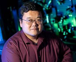
[A close up of a sign Description automatically generated] BME DISTINGUISHED RESEARCH SEMINAR SERIES November 13, 2024 9:30-10:20 am, MJIS 1001 and via ZOOM ZOOM LINK: https://us02web.zoom.us/j/89909212164?pwd=twxyjdZX8IIU3sglvspbepJ8FSZ0NC.1<https://nam04.safelinks.protection.outlook.com/?url=https%3A%2F%2Fus02web.zoom.us%2Fj%2F89909212164%3Fpwd%3DtwxyjdZX8IIU3sglvspbepJ8FSZ0NC.1&data=05%7C02%7Cbmeroundtable-list%40ecn.purdue.edu%7C1a6418d596a24640fd4108dd034c7269%7C4130bd397c53419cb1e58758d6d63f21%7C0%7C0%7C638670349181316746%7CUnknown%7CTWFpbGZsb3d8eyJFbXB0eU1hcGkiOnRydWUsIlYiOiIwLjAuMDAwMCIsIlAiOiJXaW4zMiIsIkFOIjoiTWFpbCIsIldUIjoyfQ%3D%3D%7C0%7C%7C%7C&sdata=b7P6nA6%2FYnUXe%2Be2I0xWMDzSxfKazDJQHlFxojc1uBI%3D&reserved=0> *Note: Students enrolled in the seminar course are expected to attend in person. “Ultra‐high resolution structural and molecular imaging of cells and tissues” [A person in a red shirt Description automatically generated] Fang Huang Reilly Associate Professor of Biomedical Engineering, Purdue University Abstract: Since Robert Hooke’s first description of a cell in Micrographia 350 years ago, microscopy has played an important role in understanding the rules of life. Far-field fluorescence microscopy is a powerful approach in biological and biomedical research due to its live cell compatibility and molecular specificity. A major hurdle over the last century has been the limited resolution due to the diffraction of light. Super-resolution microscopy methods, such as single-molecule localization microscopy, overcome this fundamental barrier to improve the resolution limit (250-600 nm, lateral-axial) down to a few nanometers. The development and application of SMLM majorly focus on fixed cells in thin samples and cellular structures that lie on/close to the coverslip surface and therefore, the profound impact of SMLM on biophysical, medical, and biological research has yet to fully unfold. Dr. Huang will discuss some of his team’s most recent projects which explore newly available sensors/devices such as high-speed sensors and deformable mirrors, analytical methods such as deep learning, and novel optical instrumentation to allow SMLM imaging in live cells and tissue specimens. He will show the capabilities of these new imaging systems in revealing the fine details of subcellular structures from a diverse set of biological systems including viruses, bacteria, yeasts, mammalian cells, and brain sections. Bio: The Huang lab focuses on developing novel optical imaging capacities to make significant advances in defining the structure and function of cellular constituents in biological systems. Through single molecule detection and localization, the lab develops novel super-resolution instruments, single molecule analytical methods and AI driven systems to push the envelope of spatial and temporal observation limit of biomedical research. The team demonstrate their innovative imaging systems, algorithms and computation platforms across a diverse range of biological model organisms including phages, viruses, bacteria, yeasts, mammalian cell cultures, tissues, and most recently embryos. The lab establishes and maintains strong collaborations with cell and developmental biologists, biophysicists, neuroscientists, and chemists to tackle fundamental biomedical questions in areas such as cytokinesis, epigenetics, neural circuits, immunology, and cell motility. Through these collaborations, the research team generates broad impacts to reveal disease mechanisms in Alzheimer's, autism, addiction, and cancer, and advance drug development. ~ BME Host: Krishna Jayant~ -- Bmeroundtable-list mailing list Bmeroundtable-list@ecn.purdue.edu https://engineering.purdue.edu/ECN/mailman/listinfo/bmeroundtable-list

participants (1)
-
 Gelfand, Johanna K